GENETICS
2. The structure of Genes and genomes
-
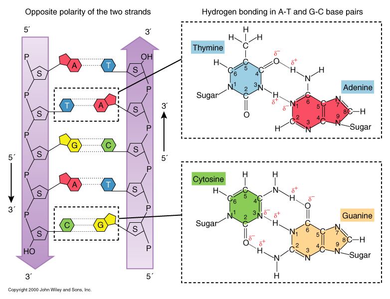
DNA is a double helix consisting of two intertwined antiparallel and complementary nucleotide chains.
-
1. 1928 Griffith: Transformation of R type Staphylococcus to S type
2. 1944 Avery, MaCarty, MacLeod: DNA as a transforming agent
3. 1950 Chargaff: [A]=[T], [G]=[C]
1950 Wilkins, Franklin: X-ray diffraction data of DNA
1953 Watson and Crick: Double helical DNA
4. The original Nature paper of Watson & Crick!!
A genome is composed of one or more DNA molecules, each organized as a chromosome.
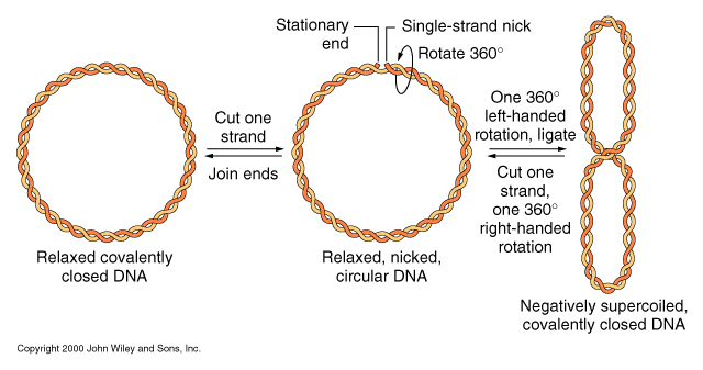
1. plasmid = mostly circular, yet a few linear DNA Prokaryotic genomes are mostly single circular chromosomes.
- mostly one circular DNA
- sometimes 2 circular DNA The nuclar complement of the genome consists of one or two sets of linear chromosomes.
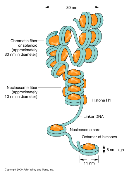
1. diverse size of each chromosome: human chromosome 1: 279 Mb, chromosome 21: 45 Mb In a nuclear chromosome DNA is wound around histone proteins, resulting in a coiled, compact, linear structure.
In a 1943 letter to his brother Roy, Oswald Avery wrote, For the past two years, first with MacLeod and now with Dr. McCarty, I have been trying to find out what is the chemical nature of the substance in the bacterial extract which induces this specific change... Some job, full of headdaches and heatbreaks. But at last perhaps we have it... But today it takes a little of well documented evidence to convince anyone that the sodium salt of deoxyribose nucleic acid, protein free, could possibly be endowed with such biologically active and specific properties, and that is the evidence we are now trying to get. It is lots of fun to blow bubbles but it is wiser to prick them yourself before someone else tries to.
Time table for revealing DNA structure and function:
See DNA structures:
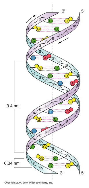
2. organellar DNA: mitochondrial and chloroplast chromosomes = circular DNA
3. viral genome = mostly linear DNA, but some linear RNA
4. prokaryotic genome
5. eukaryotic nuclear genomes
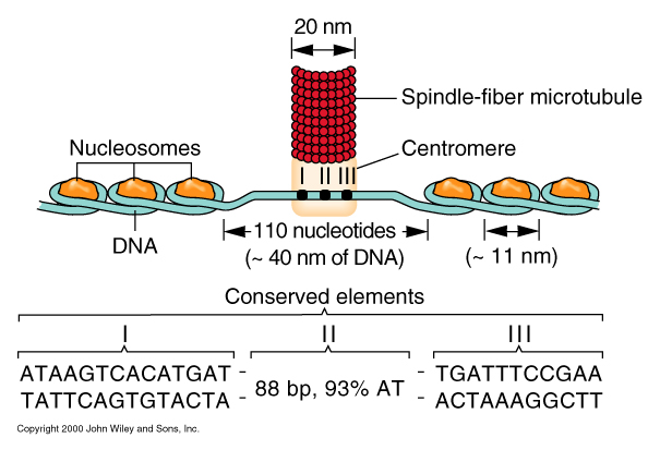
2. centromere position: metacentric (middle), acrocentric (end): Y chromosome
3. chromosome patterns: heterochromatin (highly folded region), euchromatin (less packing)
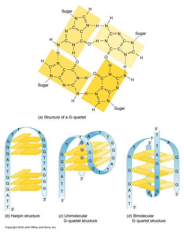
4. telomere : the end of linear DNA has a very conserved repeated sequence called as 'telomere'
For human, TTAGGG repeats up to 3,000 times in telomere.
telomeric sequence seems likely to make 'Hoogstein' base pairing: see figure (c).
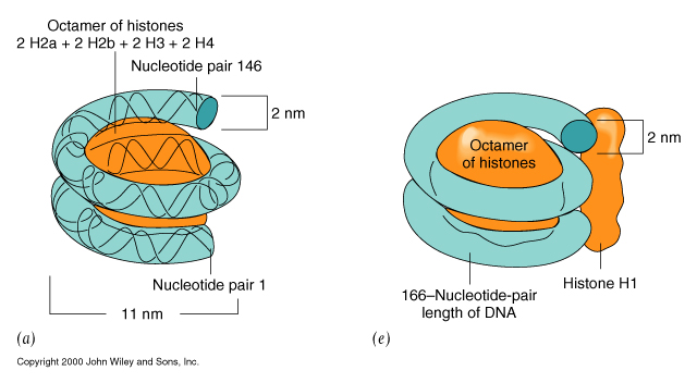
Non-dividing cells mostly contain 30 nm chromatin fiber in their nucleus.
However, metaphase chromosomes of dividing cells are densely packed: 700 nm diameter
We can see this thick chromosome with the aid of microscopes!
